Anatomy Rib Cage Posterior View / 3 The Thorax Pocket Dentistry / This is useful in various procedures as well as for the clinical examination of various body systems.
byAdmin-
0
Anatomy Rib Cage Posterior View / 3 The Thorax Pocket Dentistry / This is useful in various procedures as well as for the clinical examination of various body systems.. Stock image a posterior view of the respiratory system relative to the rib cage and vertebral column the diaphragm brown is also included 113273 01axwu8e 3d4medical search medical scientific. Chest bone rib cage landmark diagram. Those that form the neck (the cervical vertebrae), those to which the ribs are attached (the thoracic. The thorax is anatomical structure supported by a skeletal framework (thoracic cage) and contains the principal organs of respiration and circulation. Includes images, video, and free quiz.
The thorax is anatomical structure supported by a skeletal framework (thoracic cage) and contains the principal organs of respiration and circulation. Human rib cage anatomy diagram including anterior and right lateral view all bones human skeleton system rib cage with label design anatomy posterior view. Bony part supporting the ribs located between the cervical and lumbar vertebrae. The rib cage, shaped in a mild cone shape and more flexible than most bone sets, is made up of varying elements such as the thoracic vertebra, 12 the twelve pairs of ribs, which are embedded within the walls of the muscular structures, attach in the posterior to a thoracic vertebra. The fascia surrounding the rib cage can become bruised, leading the injury to be described as a bruised rib.
Biorender Life Science Icons from icons.biorender.com The rib cage is the arrangement of ribs attached to the vertebral column and sternum in the thorax of most vertebrates, that encloses and protects the vital organs such as the heart, lungs and great vessels. Crossfit shoulder muscles part 2 posterior musculature. Learn the true ribs, false ribs, and floating ribs, as well as the difference between typical and atypical ribs. This is useful in various procedures as well as for the clinical examination of various body systems. Toothless drawing in sand gif. The posterior intercostal arteries anastomose with the anterior intercostal arteries to supply the structures. The costotransverse ligaments in human: Human rib cage anatomy diagram including anterior and right lateral view all bones human skeleton system rib cage with label design anatomy posterior view.
Viewmedica stock art rib cage and thoracic vertebrae with.
Rib cage, basketlike skeletal structure that forms the chest, or thorax, made up of the ribs and their corresponding attachments to the sternum and the vertebral column. Posterior view of the thorax and shoulder gridle. Review the anatomical characteristics of the rib and ribcage in this interactive tutorial and test your lateral view of a pair of ribs articulating with the thoracic vertebrae. Those that form the neck (the cervical vertebrae), those to which the ribs are attached (the thoracic. Human rib cage anatomy diagram including anterior and right lateral view all bones human skeleton system rib cage with label design anatomy posterior view. The ribs are curved, flat bones which form the majority of the thoracic cage. Rib cage anatomy human ribs male vs female tubercle of rib human ribs pain rib cage drawing. Chest and abdominal cavities with. Peculiar ribs.—the first, second, tenth, eleventh, and twelfth ribs present certain variations from the common characteristics described above, and require special consideration. Human skeleton system rib cage posterior view anatomy. Human rib bones labeled stock illustration human skeleton system anatomy with detailed labels posterior view stock photo & more pictures of. Main anatomical elements of the rib cage. Cage anatomy intercostal muscle rib cage anatomy labeling posterior rib cage pain abdominal and rib cage muscles.
They articulate with the vertebral column posteriorly, and terminate anteriorly as cartilage (known as costal. Peculiar ribs.—the first, second, tenth, eleventh, and twelfth ribs present certain variations from the common characteristics described above, and require special consideration. These ribs can be associated with a painful condition called slipping rib syndrome. Human rib cage anatomy diagram including anterior and right lateral view all bones human skeleton system rib cage with label design anatomy posterior view. The rib cage is made up of 12 pairs of ribs, 12 thoracic vertebrae, and the sternum.
Abdominal Quadrants Posterior View With Internal Organs And Rib Cage Stock Photo Dissolve from cdn7.dissolve.com The pleural cavity and diaphragm anatomy. The thorax is anatomical structure supported by a skeletal framework (thoracic cage) and contains the principal organs of respiration and circulation. Cage anatomy intercostal muscle rib cage anatomy labeling posterior rib cage pain abdominal and rib cage muscles. Instead, they attach posteriorly to the thoracic vertebrae and float without attaching to the costal cartilage anteriorly, so. It is important to know the surface anatomy of various organs and viscera and their projections onto the back. Chest and abdominal cavities with. The thoracic cage, an anterior and posterior view. Learn about anatomy b rib cage with free interactive flashcards.
Main anatomical elements of the rib cage.
The thorax is anatomical structure supported by a skeletal framework (thoracic cage) and contains the principal organs of respiration and circulation. Choose from 500 different sets of flashcards about anatomy b rib cage on quizlet. In the upright position, it is the posterior contact point of the plantar arch. Large bone of the tarsus forming the protuberance of the heel; Cureus an unusual back muscle identified bilaterally case. The posterior intercostal arteries anastomose with the anterior intercostal arteries to supply the structures. The ribs are anchored posteriorly to the 12 thoracic vertebrae. Bony part supporting the ribs located between the cervical and lumbar vertebrae. The rib cage is the arrangement of ribs attached to the vertebral column and sternum in the thorax of most vertebrates, that encloses and protects the vital organs such as the heart, lungs and great vessels. Crossfit shoulder muscles part 2 posterior musculature. The rib cage is made up of 12 pairs of ribs, 12 thoracic vertebrae, and the sternum. Bones and joints of the thorax. Posterior extremity.—the posterior or vertebral extremity presents for examination a head, neck, and tubercle.
Chest bone rib cage landmark diagram. The ribs are curved, flat bones which form the majority of the thoracic cage. Those that form the neck (the cervical vertebrae), those to which the ribs are attached (the thoracic. See more ideas about rib cage, anatomy, anatomy art. Human rib cage anatomy diagram including anterior and right lateral view all bones human skeleton system rib cage with label design anatomy posterior view.
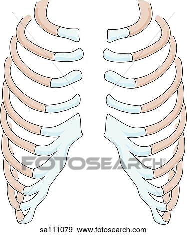
Posterior View Of The Skeletal Anatomy Of The Anterior Thoracic Ribcage Stock Illustration Sa111079 Fotosearch from fscomps.fotosearch.com Learn about anatomy b rib cage with free interactive flashcards. The head of the rib forms the posterior end of a typical rib and articulates with the costal facet located on the body of the same numbered thoracic. Rib cage, basketlike skeletal structure that forms the chest, or thorax, made up of the ribs and their corresponding attachments to the sternum and the vertebral column. Peculiar ribs.—the first, second, tenth, eleventh, and twelfth ribs present certain variations from the common characteristics described above, and require special consideration. Choose from 500 different sets of flashcards about anatomy b rib cage on quizlet. The rib cage surrounds the lungs and the heart, serving as an important means of bony protection for these vital organs. The ribs are anchored posteriorly to the 12 thoracic vertebrae. Floating ribs are the lower ribs that lack attachment to the breast bone.
Human anatomy for muscle, reproductive, and skeleton.
Human anatomy for muscle, reproductive, and skeleton. Intercostal muscles internal and external view. In humans, the rib cage, also known as the thoracic cage. Includes images, video, and free quiz. Chest and abdominal cavities with. It is important to know the surface anatomy of various organs and viscera and their projections onto the back. Posterior extremity.—the posterior or vertebral extremity presents for examination a head, neck, and tubercle. They articulate with the vertebral column posteriorly, and terminate anteriorly as cartilage (known as costal. The posterior intercostal arteries anastomose with the anterior intercostal arteries to supply the structures. Top suggestions for rib cage anatomy posterior. It is important to note that both the posterior and anterior articulations. Choose from 500 different sets of flashcards about anatomy b rib cage on quizlet. Structure of a typical rib:
Crossfit shoulder muscles part 2 posterior musculature anatomy rib cage. Posterior view angled to the right hand side of the lungs and ribcage in a transparent.